Compact Bone Diagram Lacunae - Between the rings of matrix, the bone cells (osteocytes) are located in spaces called lacunae.. (on textbook page diagrams note only highlighted labels) compact bone. Compact bone is the denser, stronger of the two types of bone tissue ( (figure) ). A structural unit of compact bone consisting central haversian canal. Look at the lacunae in the lower left corner of the image. Compact bone diagram bone cross section diagram file624 diagram of compact bone new.
The osteocytes themselves are not preserved in this type of preparation. In long bones, as you move from the outer cortical compact bone to the inner medullary cavity, the bone transitions to spongy bone. Compact bone consists of closely packed osteons or haversian systems. Noun plural lacunae luh kyoo nee ləˈkyu ni lacunas. The two tissues serve different purposes in bones, with.
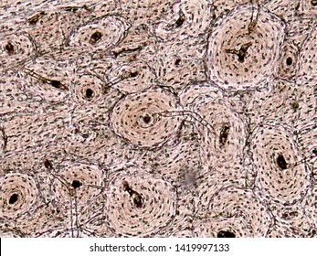
Cartilage Bone Ossification The Histology Guide from www.histology.leeds.ac.uk Due to its function, compact bone is also referred to as strong bone; There are pores and spaces even in compact bone. (the dark purple structures in the diagram above are the lacunae.) the osteocytes or mature bone cells are located in the lacunae. Haversian canals (sometimes canals of havers) are a series of microscopic tubes in the outermost region of bone called cortical bone. Learn vocabulary, terms, and more with flashcards, games, and other study tools. The lamellae are difficult to distinguish, but the haversian canals and the lacunae can be easily. Riesige auswahl an cds, vinyl und mp3s. Noun plural lacunae luh kyoo nee ləˈkyu ni lacunas.
Diagram of a typical long bone showing both cortical (compact) and cancellous (spongy) bone.
Compact bone diagram bone cross section diagram file624 diagram of compact bone new. Like compact bone, spongy bone, also known as cancellous bone, contains osteocytes housed in lacunae, but they are not arranged in concentric circles. Diagram of a typical long bone showing both cortical (compact) and cancellous (spongy) bone. To recognise bone and understand its structure and to understand the processes by which bone can be formed. Between the rings of matrix, the bone cells (osteocytes) are located in spaces called lacunae. Label the structural components of bone tissue in the diagram: Compact bone is the denser, stronger of the two types of bone tissue ( link ). The compact bones form the hard exterior of the bones, whereas the spongy bones have several pores that are filled with nerves and blood vessels. Compact bone consists of closely packed osteons or haversian systems. A diagram of the anatomy of a bone, showing the compact bone. They have long extensions that project into the canaliculi. Human bone generally comprises osseous tissue, an outer coating called a periosteum, and bone marrow.the two main structural components typically include spongy bone on the interior, with an outer layer of compact bone. Like compact bone, spongy bone, also known as cancellous bone, contains osteocytes housed in lacunae, but they are not arranged in concentric circles.
They allow blood vessels and nerves to travel through them to supply the osteocytes. I.e., slide 56, however, is not a ground section. Osteocyte is an entrapped osteoblast in the matrix. About press copyright contact us creators advertise developers terms privacy policy & safety how youtube works test new features press copyright contact us creators. Riesige auswahl an cds, vinyl und mp3s.
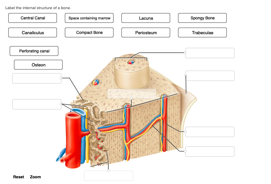
Compact Bone Tissue Images Stock Photos Vectors Shutterstock from image.shutterstock.com A diagram of the anatomy of a bone, showing the compact bone. Ultrastructure of bone components structure teachmeanatomy : Lacunae are small cavities or chambers located between one lamella and the next. Human bone generally comprises osseous tissue, an outer coating called a periosteum, and bone marrow.the two main structural components typically include spongy bone on the interior, with an outer layer of compact bone. Osteocyte is an entrapped osteoblast in the matrix. Concentric lamellae interstitial lamellae central canal lacuna osteocyte canaliculus. The dark spots are the lacunae where osteocytes would normally be found. Compact bone accounts for 80% of the bones in the human body.
I.e., slide 56, however, is not a ground section.
Learn vocabulary, terms, and more with flashcards, games, and other study tools. It can be found under the periosteum and in the diaphyses of long bones, where it provides support and protection. Diagramme schnell und einfach erstellen. Like compact bone, spongy bone, also known as cancellous bone, contains osteocytes housed in lacunae, but they are not arranged in concentric circles. A diagram of the anatomy of a bone, showing the compact bone. Learn vocabulary, terms, and more with flashcards, games, and other study tools. Canaliculus compact bone haversian canal lacuna lamellae osteocyte osteon ii spongy bone check answer. It can be remodeled all throughout life to withstand stress. About press copyright contact us creators advertise developers terms privacy policy & safety how youtube works test new features press copyright contact us creators. (the dark purple structures in the diagram above are the lacunae.) the osteocytes or mature bone cells are located in the lacunae. Compact bone, also known as cortical bone, is a denser material used to create much of the hard compact bone. Section is composed of compact bone exactly like the compact bone on slide 54. Anatomy a cavity space or depression especially in a bone containing cartilage or bone cells.
Noun plural lacunae luh kyoo nee ləˈkyu ni lacunas. The remainder is cancellous bone, which has a spongelike appearance with numerous large spaces and is found in the. Andrew kirmayer a diagram of the anatomy of a bone, showing the compact bone. Riesige auswahl an cds, vinyl und mp3s. It can be remodeled all throughout life to withstand stress.
Label The Internal Structure Of A Bone Central Canal Chegg Com from media.cheggcdn.com I.e., slide 56, however, is not a ground section. Anatomy of a long bone proximal epiphysis diaphysis distal epiphysis compact bone spongy bone medullary cavity. Osteocyte is an entrapped osteoblast in the matrix. Look at the lacunae in the lower left corner of the image. A diagram of the anatomy of a bone, showing the compact bone. Compact bone is the denser, stronger of the two types of bone tissue ( (figure) ). They allow blood vessels and nerves to travel through them to supply the osteocytes. Andrew kirmayer a diagram of the anatomy of a bone, showing the compact bone.
To know the structures of a synovial joint and a symphysis joint (intervertebral disc).
Large bone and osteon functions. Compact bone consists of closely packed osteons or haversian systems. Noun plural lacunae luh kyoo nee ləˈkyu ni lacunas. They have long extensions that project into the canaliculi. Compact bone forms the surface of all bones. Compact bone, as opposed to spongy bone, is made of cylindrical units, called osteons, that are tightly formed together. In long bones, as you move from the outer cortical compact bone to the inner medullary cavity, the bone transitions to spongy bone. To know the structures of a synovial joint and a symphysis joint (intervertebral disc). Compact bone, also known as cortical bone, is a denser material used to create much of the hard compact bone. Like compact bone, spongy bone, also known as cancellous bone, contains osteocytes housed in lacunae, but they are not arranged in concentric circles. I.e., slide 56, however, is not a ground section. Start studying compact bone labeling. Ultrastructure of bone components structure teachmeanatomy :
Concentric lamellae interstitial lamellae central canal lacuna osteocyte canaliculus compact bone diagram. Large bone and osteon functions.


0 Komentar